Neuroimaging Labs Research Center
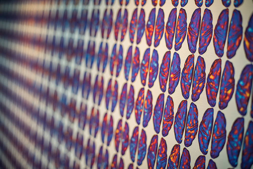
The nationally recognized Neuroimaging Labs Research Center (NIL-RC) is home to a large number of faculty, students, postdocs and staff from multiple Washington University departments. Using neuroimaging techniques and analyses largely developed over the years in the NIL-RC, investigators perform groundbreaking work in human cognitive and clinical neuroscience, human aging and neurodevelopment, human systems neuroscience, human neuropathophysiology, basic human brain physiology, and animal models of neurological disorders.
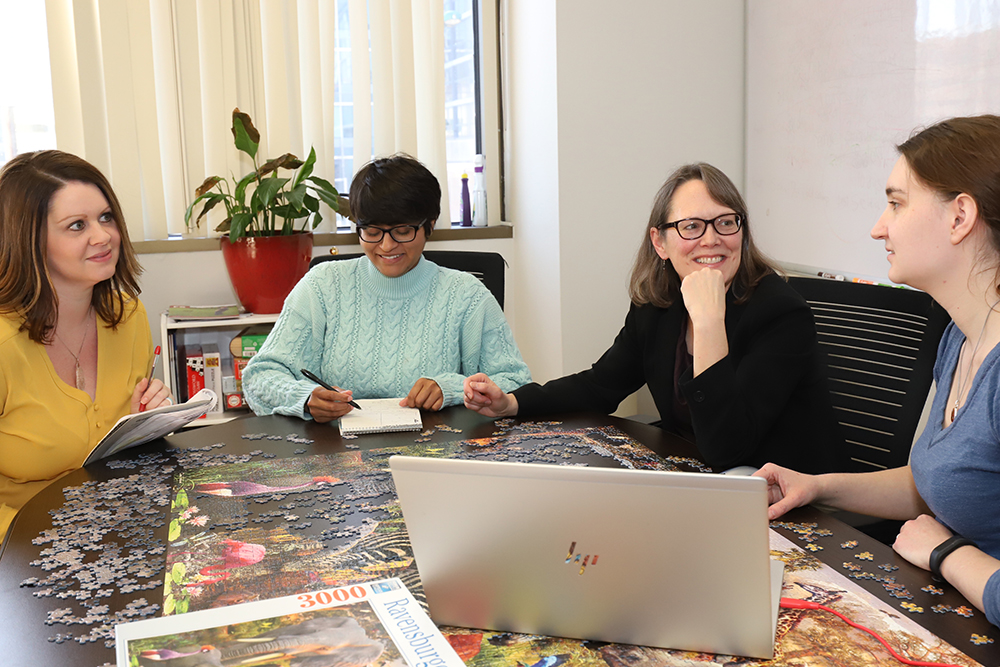
Our People
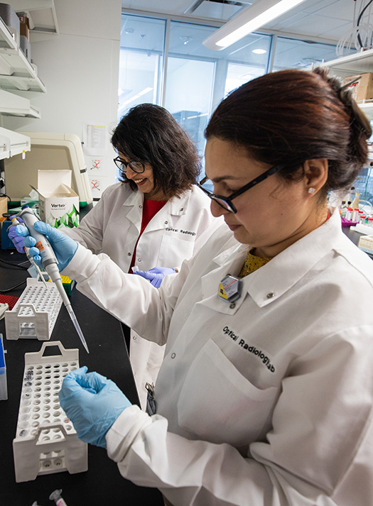
The NIL-RC provides a physical and intellectual environment that promotes collaborative, innovative and interdisciplinary neuroimaging research and supports the career development of students and junior faculty.
Our Research
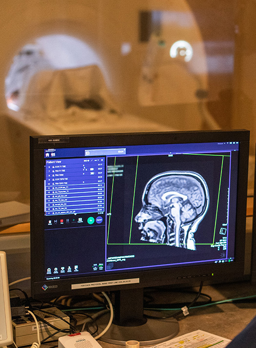
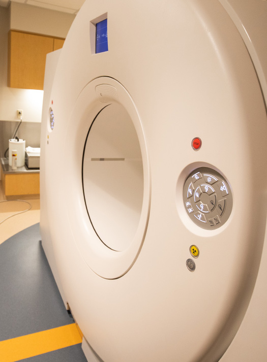
NIL-RC studies employ PET, MRI, TMS, Diffuse Optical Tomography (DOT) and EEG in conjunction with detailed behavioral analyses to understand the human and animal brain in health and disease.
Group Resources
The NIL-RC has provided valuable support for many kinds of other large-scale initiatives including the Human Connectome Project and Alzheimer’s Disease Research Center.